
“Even traffic can ruin our images. That’s why the spot in the middle of campus is ideal”

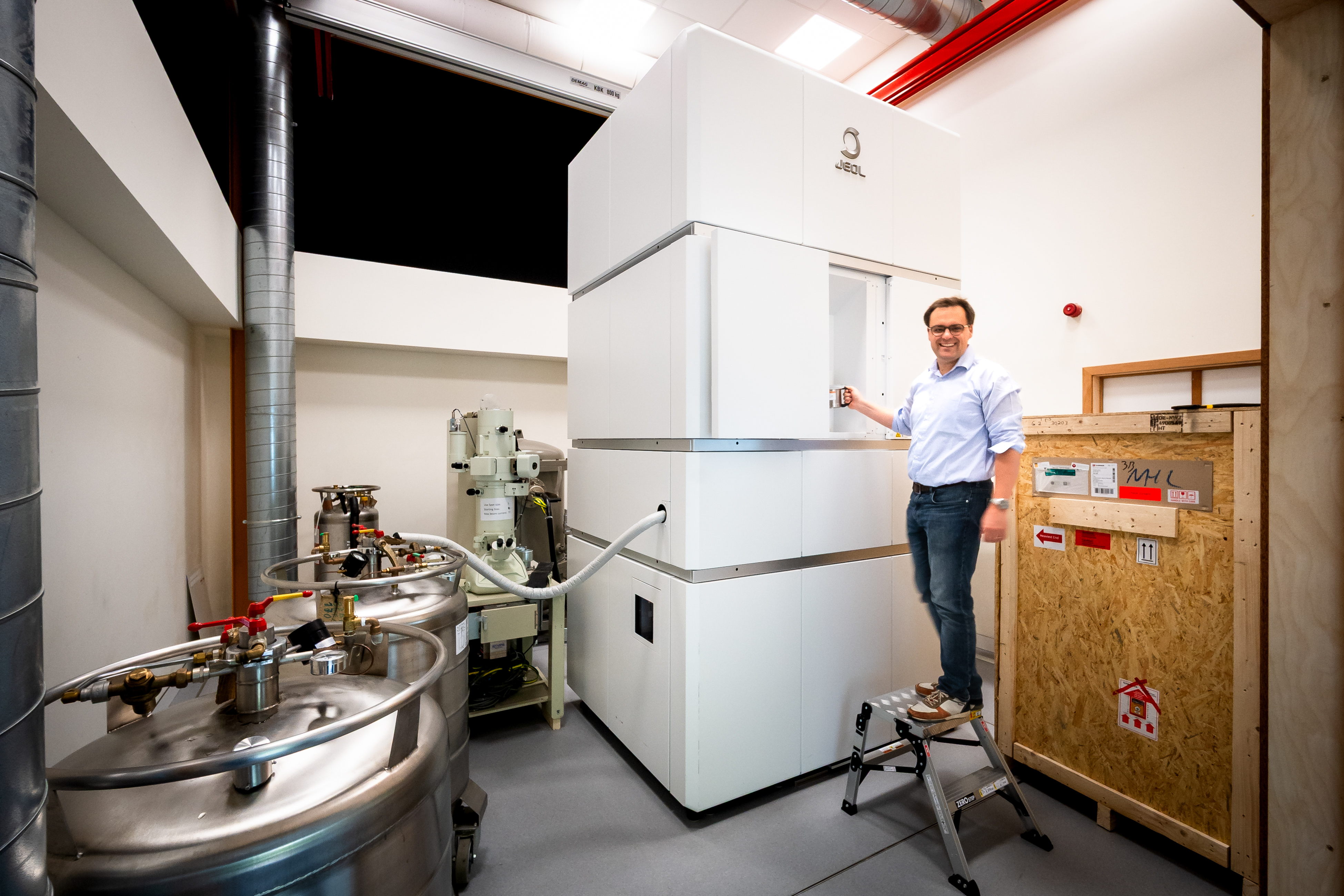
In the heart of the VUB campus stands the Electron Cryogenic Microscope. This unique (and extremely costly) giant microscope can capture the three-dimensional structure of proteins – atom by atom. The CRYO ARM300 is used mainly for fundamental research, but also plays a role in the search for new medicines. Its location is just as remarkable: Building P1, once the student halls designed by Willy Van Der Meeren.
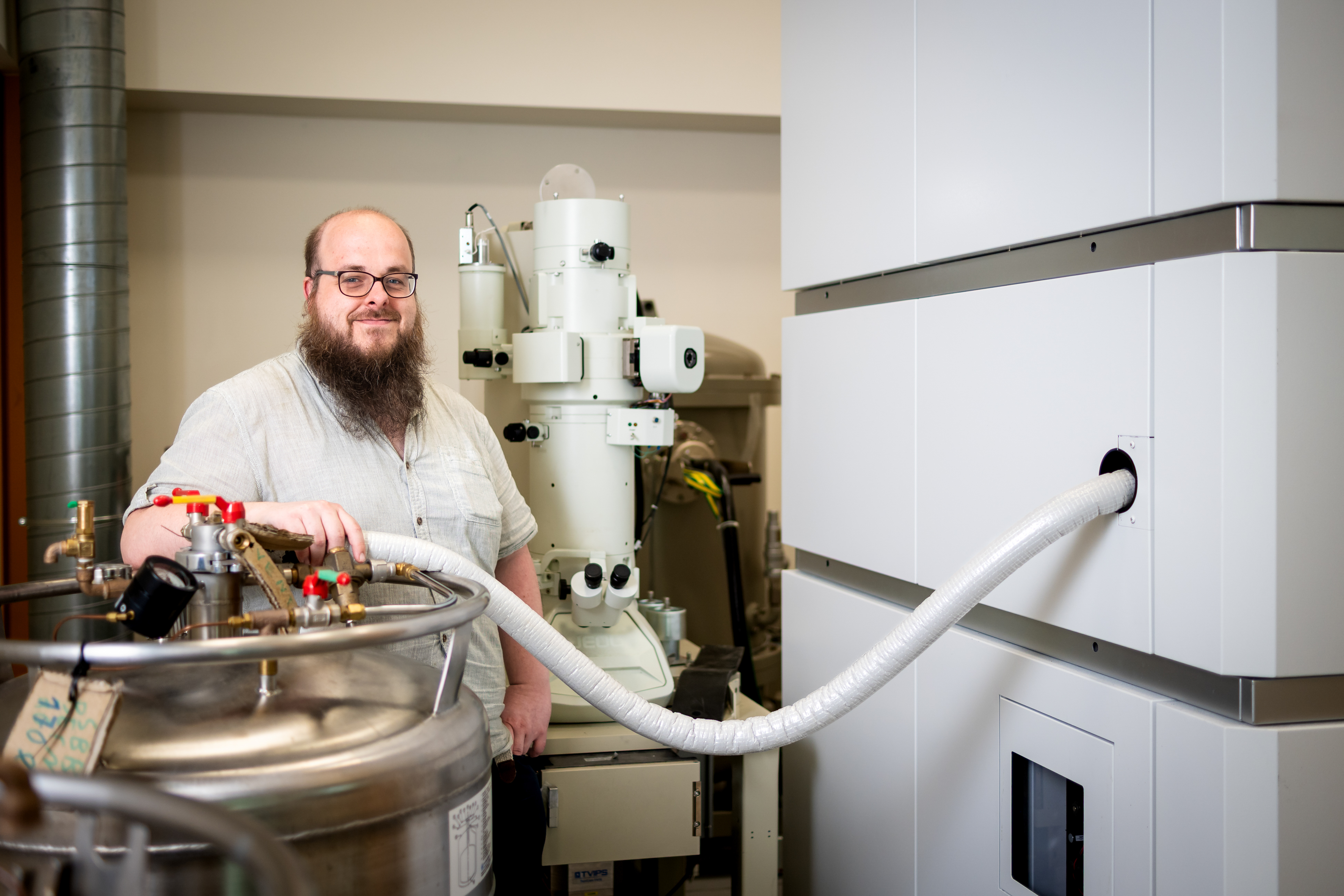
A maze of cardboard boxes and spare parts, with a racing bike somehow squeezed in between: the office of Marcus Fislage, facility manager of the Centre for Bio Electron Cryogenic Microscopy (BECM), has all the charm of Boys & Science. The same goes for the giant microscope at the core of this research centre. When the CRYO ARM300 (price tag: four million euros) was installed here in 2018, it was – together with Glasgow – the first of its kind outside Japan, the country where these machines are built. But how did such a piece of high-tech kit end up in a building once buzzing with students enjoying their first taste of independence?


Professor Rouslan Efremov (VIB-VUB Centre for Structural Biology): “We needed a space with sufficient height – the microscope is over three metres tall. Willy Van Der Meeren’s student halls turned out to be perfect. They are prefab constructions that can be easily adapted for other purposes. In this case, the ceiling between two rooms was removed to create the height we needed. The other rooms of the red cluster have been converted into our offices and laboratories.”
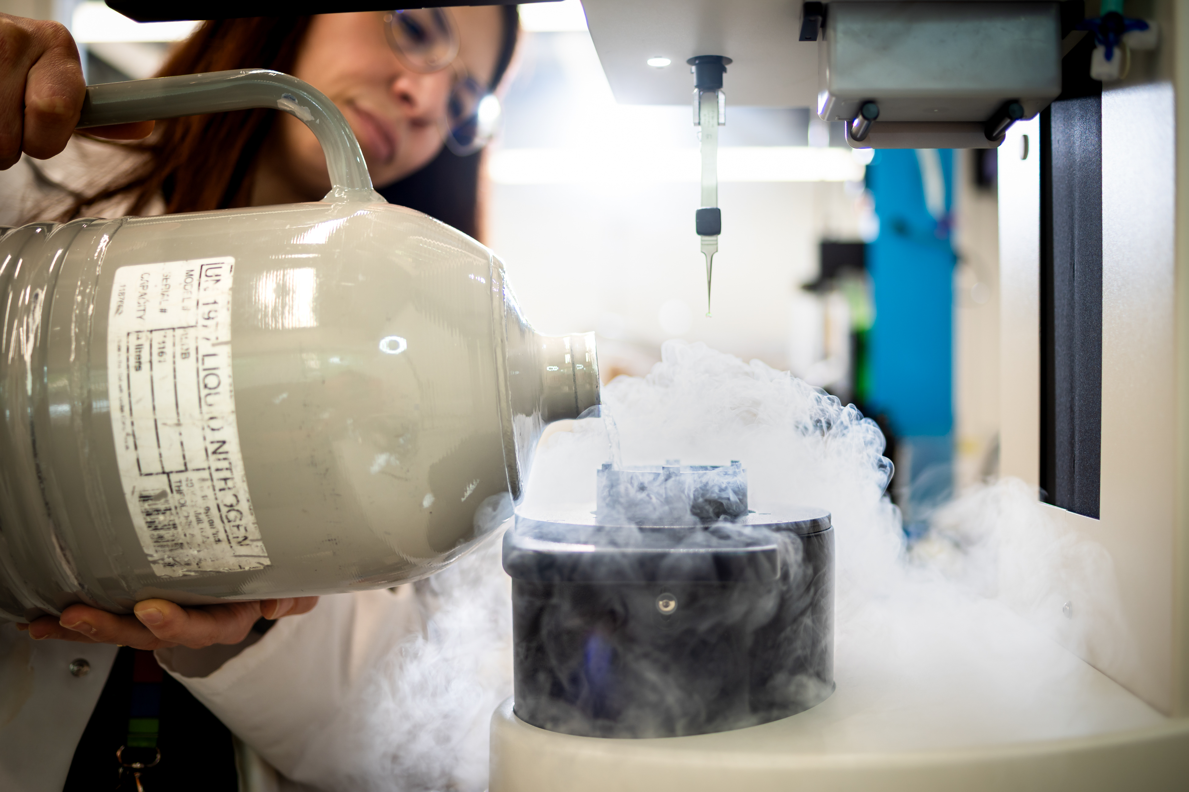
In one of these labs, proteins are prepared before being placed under the microscope. A researcher puts a purified protein sample onto a tiny grid. This is then plunged into liquid ethane in a so-called plunge freezer. Within milliseconds, the sample is frozen to -200°C.
Rouslan Efremov: “This ultra-fast freezing has two advantages: the proteins keep their natural shape, and no ice crystals form in the frozen water. The result is a very thin, perfectly transparent layer of ice with intact proteins trapped inside. This frozen protein sample is then placed in the cryo-electron microscope and exposed to an electron beam from above. A camera underneath detects the electrons that pass through and produces thousands of two-dimensional images for each sample. Specialised software processes and combines these into a high-resolution 3D reconstruction of the protein structure – including the nitrogen, oxygen and carbon molecules of the amino acids, the building blocks of proteins.”
Marcus Fislage: “For many scientists, this is an incredibly moving moment. Some even get emotional. They may have been studying a particular protein for years, but this is the first time they can actually see it in 3D.”
"The slightest vibration affects the microscope and the quality of the images"
Cryogenic microscopy is not a matter of millimetres, but of nanometres – even Angstroms, ten times smaller than a nanometre. In other words: atomic level. The tiniest vibration can affect the functioning of the microscope and the quality of the images.
Marcus Fislage: “The VUB campus is surrounded by cars, trams and trains. That’s great for visiting scientists, who can get here easily by public transport and also use the campus restaurants – handy, as they usually spend at least a full day here. But we still had to eliminate the vibrations.”
It helps that the Van Der Meeren halls stand in the middle of campus, away from the traffic. But that alone is not enough. Even a slamming door or a shouting student could disturb the microscope.
Marcus Fislage: “That’s why the foundation beneath the equipment consists of a three-metre-thick layer of concrete. That, in turn, rests on steel pillars driven six metres into the ground. The room is also acoustically very well insulated, and we have a system in place that captures and neutralises electromagnetic signals from passing trains before they can enter my lab.”
‘My room’? “Yes, I take my job as facility manager very seriously. (laughs) I also call them ‘my’ microscopes. We have three here: the CRYO ARM300 and two smaller ones used to test the quality of protein samples. We even gave them names: Kathleen, Karen and Kristel.”
Rouslan Efremov: “The camera in the microscope is a K3, and my children were fans of the girl band, hence the names.”
Marcus Fislage: “Right now we’re building a fourth microscope – a smaller version of the CRYO ARM300. It will take over some of the more routine tasks from the big one, but it’s much more compact and costs only one million instead of four. We still need to think of a name for that one…”
“The better you understand a protein’s structure and function, the more targeted your search for medicines becomes”
Jokes aside, Marcus has serious responsibilities as facility manager. He oversees maintenance and repairs (“these are ultra-sophisticated devices, so things break easily”), and supports the scientists who use the infrastructure.
Marcus Fislage: “The researchers come from VUB, but also from other Belgian and sometimes foreign universities. Companies too – such as the VUB spin-off Confo Therapeutics – regularly make use of the BECM. It makes sense to share expensive equipment as efficiently as possible. The microscope is available 24/7.”

The BECM is used mainly for fundamental research, but the step towards medical applications is quickly taken. Each protein has a unique structure and a specific role in the cell. Many diseases arise when something goes wrong at this level. Medicines can correct this. They bind to the protein molecules that cause a disease and influence their function. For example, Prozac blocks the reuptake of serotonin by binding to transport proteins. ACE inhibitors bind to the angiotensin-converting enzyme – another protein – and lower blood pressure.
Rouslan Efremov: “The better you understand a protein’s structure and function, the more targeted your search for new medicines can be. Pharmaceutical companies have many candidate molecules that could work against various diseases. Thanks in part to the BECM, they can now test and identify effective ones faster than ever before.”
Marcus Fislage: “The images from the BECM are incredibly detailed and of exceptional quality, and the software to analyse them is more powerful than ever. The difference compared to ten years ago is immense. Back then, this was still science fiction."
Cracking Alzheimer’s at atomic level
Ruslan Efremov is Associate Professor at the Vrije Universiteit Brussel and group leader within Structural Biology Brussels. His research focuses on the structure and function of biological molecular machines, such as protein complexes that play a crucial role in health and disease. Using advanced cryo-electron microscopy, he visualises these complex structures at atomic resolution. His work contributes to a deeper understanding of fundamental biological processes and supports the development of new biomedical applications.

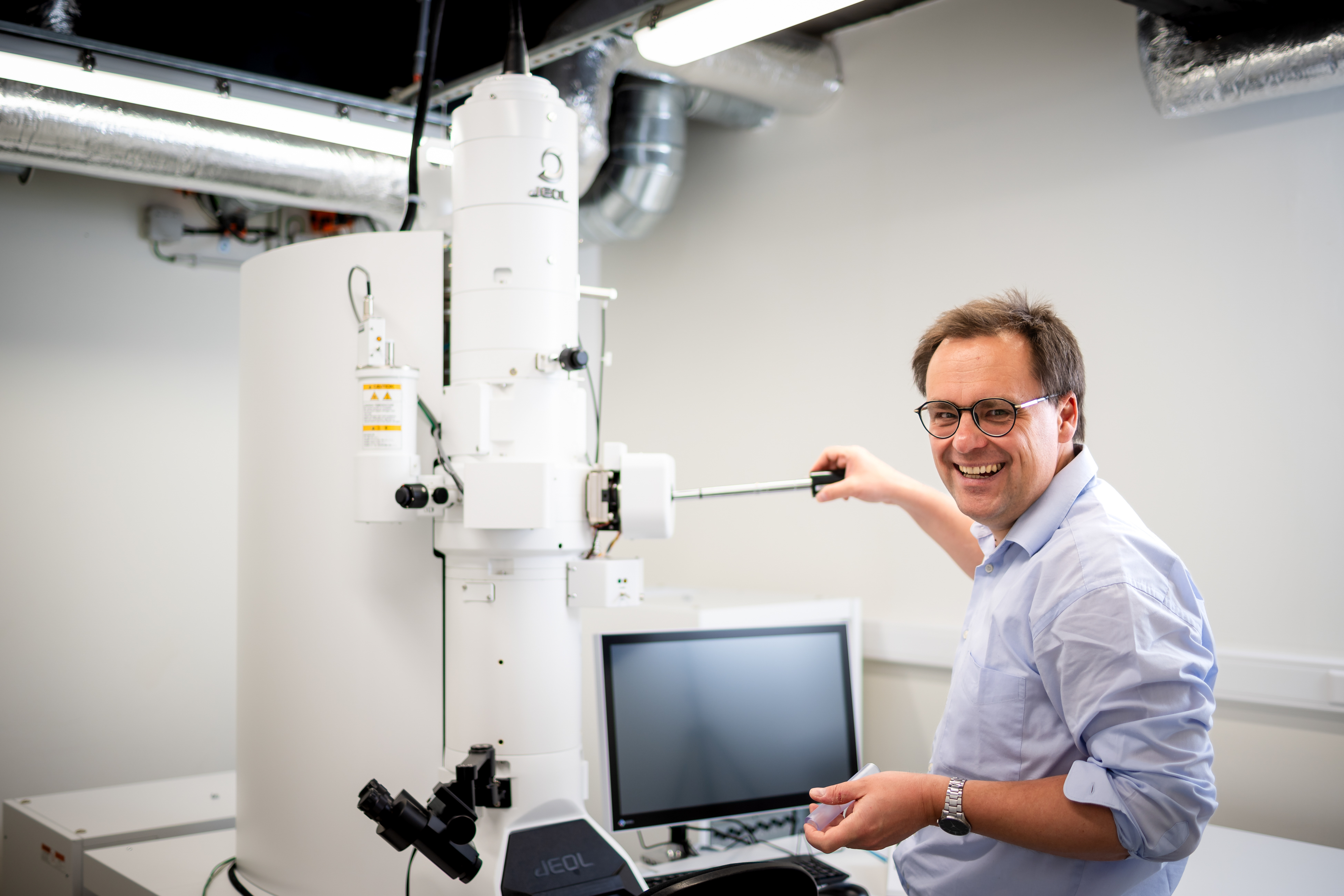
Marcus Fislage is a postdoctoral researcher at Structural Biology Brussels (VUB) and manager of the VUB-VIB cryo-electron microscopy facility. His research focuses on the structure and function of biomolecular machines that are essential to fundamental cellular processes. Using advanced cryo-EM technology, he characterises protein complexes and RNA-related mechanisms at high resolution. His work provides important insights into the molecular basis of health and disease and supports innovation in biomedical research.
In this article:
- How does a four-million-euro microscope make the invisible visible?
- Why must proteins be frozen within a fraction of a second?
- How does a detailed 3D image of proteins support the development of new medicines?
- Why can even a slamming door or passing train disrupt an experiment?


